Ventricle in newborns. Enlargement of the ventricles of the brain: consequences of enlargement and asymmetry in newborns and infants
Neurosonography (NSG) is a term applied to the study of the brain of a young child: a newborn and an infant until the fontanel closes using ultrasound.
Neurosonography, or ultrasound of the child’s brain, can be prescribed by a pediatrician at the maternity hospital or a neurologist at a children’s clinic in the 1st month of life as part of screening. In the future, according to indications, it is carried out on the 3rd month, on the 6th month and until the fontanelle closes.
As a procedure, neurosonography (ultrasound) is one of the safest research methods, but it should be carried out strictly as prescribed by the doctor, because Ultrasonic waves can have a thermal effect on body tissue.
At the moment, no negative consequences from the neurosonography procedure have been identified in children. The examination itself does not take much time and lasts up to 10 minutes, and it is completely painless. Timely neurosonography can save the health and sometimes even the life of a child.
Indications for neurosonography
The reasons for requiring ultrasound scanning in the maternity hospital are varied. The main ones are:
- fetal hypoxia;
- asphyxia of newborns;
- difficult labor (accelerated/prolonged, with the use of obstetric aids);
- intrauterine fetal infection;
- birth injuries of newborns;
- infectious diseases of the mother during pregnancy;
- Rhesus conflict;
- C-section;
- examination of premature newborns;
- detection of fetal pathology on ultrasound during pregnancy;
- less than 7 points on the Apgar scale in the delivery room;
- retraction/protrusion of the fontanelle in newborns;
- suspicion of chromosomal pathologies (according to a screening study during pregnancy).
 The birth of a child by cesarean section, despite its prevalence, is quite traumatic for the baby. Therefore, children with such a history are required to undergo NSG for early diagnosis of possible pathology
 Indications for ultrasound examination within a month:
- suspicion of ICP;
- congenital Apert syndrome;
- with epileptiform activity (NSH is an additional method for diagnosing the head);
- signs of strabismus and diagnosis of cerebral palsy;
- head circumference is not normal (symptoms of hydrocephalus/dropsy);
- hyperactivity syndrome;
- injuries to the child's head;
- delay in the development of the infant's psychomotor skills;
- sepsis;
- cerebral ischemia;
- infectious diseases (meningitis, encephalitis, etc.);
- rickety shape of the body and head;
- CNS disorders due to a viral infection;
- suspicion of neoplasms (cyst, tumor);
- genetic developmental abnormalities;
- monitoring the condition of premature babies, etc.

 In addition to the main causes, which are serious pathological conditions, NSG is prescribed when a child’s fever lasts for more than a month and has no obvious cause
 Preparation and method of conducting the study
Neurosonography does not require preliminary preparation. The baby should not be hungry or thirsty. If the baby falls asleep, there is no need to wake him up; this is even welcome: it is easier to ensure that the head remains still. The results of neurosonography are issued 1-2 minutes after the completion of the ultrasound.
You can take baby milk and a diaper with you to place your newborn baby on the couch. Before the NSG procedure, there is no need to apply creams or ointments to the fontanel area, even if there are indications for this. This worsens the contact of the sensor with the skin and also negatively affects the visualization of the organ being studied.
The procedure is no different from any ultrasound. A newborn or infant is placed on a couch, the place where the skin comes into contact with the sensor is lubricated with a special gel substance, after which the doctor performs neurosonorgraphy.
Access to brain structures with ultrasound is possible through the large fontanelle, thin temple bone, antero- and posterolateral fontanelles, as well as the foramen magnum. In a child born at term, the small lateral fontanelles are closed, but the bone is thin and permeable to ultrasound. Interpretation of neurosonography data is carried out by a qualified physician.
Normal NSG results and interpretation
Interpretation of diagnostic results consists of describing certain structures, their symmetry and echogenicity of tissues. Normally, in a child of any age, the brain structures should be symmetrical, homogeneous, and have appropriate echogenicity. In the neurosonography transcript, the doctor describes:
- symmetry of brain structures - symmetrical/asymmetrical;
- visualization of grooves and convolutions (must be clearly visualized);
- condition, shape and location of the cerebellar structures (tentory);
- condition of the medullary falx (thin hyperechoic stripe);
- presence/absence of fluid in the interhemispheric fissure (fluid should be absent);
- homogeneity/heterogeneity and symmetry/asymmetry of the ventricles;
- condition of the cerebellar tentorium (tent);
- absence/presence of formations (cyst, tumor, developmental anomaly, change in the structure of the brain matter, hematoma, fluid, etc.);
- the state of the vascular bundles (normally they are hyperechoic).
Table with standards for neurosonography indicators from 0 to 3 months:
| Options | Norms for newborns | Norms at 3 months |
|---|---|---|
| Lateral ventricles of the brain | Anterior horns – 2-4 mm. Occipital horns – 10-15 mm. Body – up to 4 mm. | Anterior horns – up to 4 mm. Occipital horns – up to 15 mm. Body – 2-4 mm. |
| III ventricle | 3-5 mm. | Up to 5 mm. |
| IV ventricle | Up to 4 mm. | Up to 4 mm. |
| Interhemispheric fissure | 3-4 mm. | 3-4 mm. |
| Large tank | Up to 10 mm. | Up to 6 mm. |
| Subarachnoid space | Up to 3 mm. | Up to 3 mm. |
The structures should not contain inclusions (cyst, tumor, fluid), ischemic foci, hematomas, developmental anomalies, etc. The transcript also contains the dimensions of the described brain structures. At the age of 3 months, the doctor pays more attention to describing those indicators that should normally change.
Pathologies detected using neurosonography
Based on the results of neurosonography, a specialist can identify possible developmental disorders of the baby, as well as pathological processes: neoplasms, hematomas, cysts:
- Choroid plexus cyst (does not require intervention, asymptomatic), usually there are several of them. These are small bubble formations containing liquid - liquor. Self-dissolving.
- Subependymal cysts. Formations whose contents are liquid. They occur as a result of hemorrhage and can occur pre- and postpartum. Such cysts require observation and, possibly, treatment, since they can increase in size (due to failure to eliminate the causes that caused them, which may be hemorrhage or ischemia).
- Arachnoid cyst (arachnoid membrane). They require treatment, observation by a neurologist and control. They can be located anywhere in the arachnoid membrane, can grow, and are cavities containing liquid. Self-resorption does not occur.
- Hydrocephalus/dropsy of the brain is a lesion that results in dilatation of the ventricles of the brain, as a result of which fluid accumulates in them. This condition requires treatment, observation, and control of the NSG over the course of the disease.
- Ischemic lesions also require mandatory therapy and dynamic control studies using NSG.
- Hematomas of brain tissue, hemorrhages into the ventricular space. Diagnosed in premature babies. In full-term infants, this is an alarming symptom and requires mandatory treatment, monitoring and observation.
- Hypertension syndrome is, in fact, an increase in intracranial pressure. It is a very alarming sign of a significant shift in the position of any hemisphere, both in premature and full-term babies. This occurs under the influence of foreign formations - cysts, tumors, hematomas. However, in most cases, this syndrome is associated with an excess amount of accumulated fluid (CSF) in the brain space.
If any pathology is detected by ultrasound, you should contact special centers. This will help you get qualified advice, make a correct diagnosis and prescribe the correct treatment regimen for your child.
The ventricles of the brain are a system of cavities connected by channels. A fluid circulates in these spaces - cerebrospinal fluid. It nourishes the tissue of the nervous system and ensures the outflow of metabolic products.
When exposed to negative factors, a pathology is formed - expansion of the ventricles of the brain. Most often it is detected in newborns during the first comprehensive examination of the nervous system.
It should be remembered that not every increase in the size of the ventricles is a pathology. An anomaly is considered a disease if it causes symptoms, disrupts the body’s adaptation and worsens a person’s quality of life.
Enlargement of the ventricles of the brain is formed due to the influence of such factors:
- Skull trauma at birth. This happens if the mother's birth canal does not match the size of the fetus's head. For example, if the mother has a narrow pelvis and the child has a large head circumference.
- Congenital anatomical features. Some have long fingers, some have big ears, some have wide ventricles in the brain.
- Violation of the outflow of cerebrospinal fluid, resulting in an excess of fluid in the cavities. This is observed in diseases accompanied by mechanical compression of the cerebrospinal fluid ducts. For example, a tumor in the cerebral hemispheres or a herniation of the spinal cord.
- In adults, ventriculomegaly develops as a result of hemorrhagic stroke - an acute circulatory disorder in which blood enters the brain and can enter the ventricles.

Symptoms and manifestations
Enlargement and dilation of the ventricles of the brain can occur as hydrocephalus and hypertensive-hydrocephalic syndrome.
Characteristics of the first type.
According to the clinical picture, hydrocephalus differs in children under one year of age and children after one year of age. In the first option, the shape and size of the infant's head changes: the forehead protrudes above the face. The scalp turns pale and wrinkles, becoming like the head of old people.
With hydrocephalus in children after one year, a progressive change in the sutures is observed.
Symptoms of ventricular enlargement are caused by increased intracranial pressure. In parallel, atrophic and degenerative changes develop in the central nervous system.
In newborn babies, the eyes shift downwards - this is a symptom of the “setting sun”. Accuracy decreases and fields of view become narrower. Pathology can lead to complete or partial loss of vision. The abducens nerve is affected. This leads to squint. Movement disorders develop: paresis - weakening of the strength of skeletal muscles.
The cerebellum is affected. Coordination and statics are upset. As a rule, hydrocephalic children are severely retarded in intellectual and physical development. Their emotional sphere is disturbed: they are irritable, excitable, or vice versa, often lethargic and apathetic. They do not play with other children and have difficult contact with them.
Hydrocephalus in adolescents and adults is manifested by severe morning headache, nausea and vomiting. In patients, the functions of higher nervous activity are inhibited. Consciousness is impaired, memory and thinking are disrupted, and speech is impaired. Patients experience swelling of the optic discs, which leads to optic nerve atrophy and vision loss.

A complication of hydrocephalus is occlusive crisis. Its cause is a sudden disruption of cerebrospinal fluid from the ventricles of the brain. The pathological condition develops quickly. The accumulated fluid compresses the brain and stem structures.
Dilatation of the 4th ventricle of the brain is the main factor in the development of occlusive crisis. In this case, the cerebrospinal fluid compresses the rhomboid fossa and the midbrain. Symptoms: acute headache, vomiting and nausea, mental agitation, forced head position. Consciousness is impaired, oculomotor functions are disrupted. In an acute condition, the autonomic nervous system is involved: sweating increases, the heart rate slows down, the skin turns pale, the face turns red and warms. Motor disturbances develop rapidly: tonic convulsions appear.
The expansion of the lateral ventricles also develops according to the second option: according to the type of hypertensive-hydrocephalic syndrome. Its signs:
- The child sucks poorly and often refuses food. If you manage to feed, the baby vomits like a fountain.
- Decreased muscle tone.
- Congenital basal reflexes are partially suppressed. Weak grasping and swallowing.
- Periodic convulsions, trembling of limbs.
- Strabismus, deterioration of visual acuity, loss of lateral fields.
- "Rising sun" symptom.
- Protruding cranial sutures.
- Rapid head growth.
In school-age children, the syndrome is usually triggered by traumatic brain injury.
What is the standard size
Normal ventricle sizes:
- The third ventricle of the brain is dilated if its dimensions exceed 5 mm.
- The depth of the fourth cavity is no more than 4 mm.
- The depth of the lateral ventricles is no more than 4 mm.
Diagnosis and treatment
Diagnosis of dilated ventricles is carried out on the basis of signs and results of instrumental and additional examination methods. The main thing is to observe the child in dynamics. The doctor is interested in the functionality of higher nervous activity, the behavior and mental sphere of the child, accuracy of vision, coordination and the presence of seizures.
Instrumental research methods:
- Ventriculography.
- Pneumoencephalography.
The most effective way to quickly diagnose ventricular enlargement is to carry out. It can be carried out even during the mother’s pregnancy.
Treatment is aimed at reducing intracranial pressure and allowing cerebrospinal fluid to pass through. For this, diuretics are prescribed. They also give medications that improve blood flow to the brain.
The human brain is a complex and amazing structure, all the mysteries of which scientists have not yet unraveled. One of the most interesting mechanisms of the functioning of the nervous system remains the process of formation and circulation of cerebrospinal fluid (CSF), which is carried out using the 3rd ventricle of the brain.
3rd ventricle of the brain: anatomy and physiology
The third ventricle of the brain is a thin slit-like cavity bounded by the visual thalamus and located in the diencephalon. Inside, the third ventricle of the brain is lined with a soft membrane, a branched choroid plexus and filled with cerebrospinal fluid.
The physiological significance of the 3rd ventricle is very great. It ensures unimpeded flow of cerebrospinal fluid from the lateral ventricles into the subarachnoid space for washing the brain and spinal cord. Simply put, it ensures the circulation of cerebrospinal fluid, which is necessary for:
- regulation of intracranial pressure;
- mechanical protection of the brain from damage and injury;
- transporting substances from the brain to the spinal cord and vice versa;
- protecting the brain from infection.
3rd ventricle of the brain: normal in children and adults
A normally functioning liquor system is an uninterrupted and harmonious process. But if even a small “breakdown” occurs in the processes of formation and circulation of cerebrospinal fluid, this will certainly affect the condition of the child or adult.
The 3rd ventricle of the brain is especially important in this regard, the norm of which is indicated below:
- Newborns -3-5 mm.
- Children 1-3 months -3-5 mm.
- Children 3 months - 6 years -3-6 mm.
- Adults -4-6 mm.
Common diseases of the third ventricle of the brain
Most often, the problem of impaired outflow of cerebrospinal fluid occurs in children - newborns and babies up to one year old. One of the most common diseases at this age is ICH () and its complication – hydrocephalus.
During pregnancy, the expectant mother undergoes mandatory ultrasound examinations of the fetus, which make it possible to identify congenital malformations of the child’s central nervous system in the early stages. If during the examination the doctor notes that the 3rd ventricle of the brain is dilated, additional diagnostic tests and careful medical supervision will be needed.
If the cavity of the 3rd ventricle in the fetus becomes more and more dilated, in the future such a baby may require bypass surgery to restore the normal outflow of cerebrospinal fluid.
Also, all born babies at two months of age (earlier if indicated) undergo a mandatory medical examination by a neurologist, who may suspect dilation of the 3rd ventricle and the presence of ICH. Such children are sent for a special examination of brain structures (neurosonogathia).
What is NSG?
Neurosonography is a special type of ultrasound examination of the brain. It can be performed on infants because they have a small physiological opening in the skull - the fontanel.
Using a special sensor, the doctor receives an image of all the internal structures of the brain, determines their size and location. If the 3rd ventricle is dilated in the NSG, more detailed tests are performed - computed tomography (CT) or magnetic resonance imaging (MRI) to obtain a more accurate picture of the disease and confirm the diagnosis.
Which doctors should you contact when diagnosing ICH?
If the 3rd ventricle of the baby’s brain is slightly enlarged and the mother has no serious complaints, regular monitoring by a local pediatrician is sufficient. Consultation with a neurologist and neurosurgeon is necessary if there is significant dilatation of the ventricles on ultrasound or symptoms of ICH:
- the child began to suck the breast worse;
- the fontanel is tense, protruding above the surface of the skull;
- the saphenous veins of the scalp are dilated;
- Graefe's symptom - a section of white sclera between the iris and eyelid when looking down;
- loud, sharp cry;
- vomit;
- divergence of the sutures of the skull;
- rapid increase in head size.
Doctors determine further tactics for treating a baby with: conservative means prescribing vascular medications, massage, physiotherapy; surgical – performing an operation. After therapy, children quickly recover, the activity of the nervous system is restored.
 Colloid cyst of the 3rd ventricle is a disease common among adults 20-40 years old. It is characterized by the appearance of a benign round formation in the cavity of the 3rd ventricle, which is not prone to rapid growth and metastasis.
The colloid cyst itself does not pose any danger to human health. Problems begin if it reaches a large size and interferes with the outflow of cerebrospinal fluid. In this case, the patient experiences neurological symptoms associated with increased intracranial pressure:
- severe headache;
- vomit;
- visual impairment;
- convulsions.
Diagnosis and treatment of colloid cyst of the third ventricle are jointly carried out by a neurologist and a neurosurgeon. If the size of the formation is pronounced, determined on CT or, surgical treatment of the cyst is prescribed. After the operation, the normal flow of cerebrospinal fluid is quickly restored, and all symptoms of the disease disappear.
Summing up
Thus, the third ventricle is an important element of the cerebrospinal fluid system, diseases of which can lead to serious consequences. Attentive attention to health and timely consultation with doctors will help you quickly and permanently cope with the disease.
In this case, health assessment is carried out in stages, starting from the first minute of life, and ends before discharge.
The most thorough examination is carried out on the first day and consists of a standard procedure for monitoring the activity and appearance of the newborn. If the doctor suspects congenital malformations, then it is possible to use an ultrasound examination, which can reveal abnormalities in the formation of not only internal organs, but also the brain. In this case, the sizes of the ventricles are especially carefully measured, which normally should not exceed a certain value.
At this stage, the neonatologist can diagnose dilation of the ventricles of the brain in newborns. Based on the degree of pathology and the impact on the child’s life, the question will be raised about further solving this problem: for example, in case of minor deviations from the norm, observation by a neurologist and monitoring of the condition are prescribed. If the violations are serious and the symptoms are pronounced, then the child needs special treatment and observation in a hospital setting.
The ventricular system consists of 4 cavities located in parts of the brain. Their main purpose is the synthesis of cerebrospinal fluid or cerebrospinal fluid, which performs a large number of tasks, but its main function is to cushion the brain matter from external influences, control intracranial pressure and stabilize metabolic processes between the blood and the brain.
The movement of cerebrospinal fluid occurs through channels connecting the common 4th ventricle and the subarachnoid space formed by the membranes of the spinal cord and brain. Moreover, its main volume is located above significant fissures and convolutions of the cortex.
The largest lateral ventricles are located equidistant from the midline below the corpus callosum. The first ventricle is considered to be the cavity located on the left side, and the second - on the right. They are C-shaped and wrap around the dorsal parts of the basal ganglia. They produce cerebrospinal fluid, which enters the third ventricle through the intergastric openings. Structurally, segments I and II of the ventricular system include the anterior (frontal) horns, body and inferior (temporal) horns.
The third ventricle is located between the visual tuberosities and has the shape of a ring. At the same time, gray matter is located in its walls, which is responsible for regulating the autonomic system. This section is connected with the midbrain aqueduct, and through the interventricular foramen, located behind the nasal commissure, with the I and II ventricles.
The most important IV ventricle is located between the cerebellum and the medulla oblongata, with the vermis and medullary velum located above it, and the medulla oblongata and pons below it. This cavity was formed from the remains of the posterior medullary vesicle and is common to the rhomboid region. At its bottom lie the nuclei of the V-XII cranial nerves. In this case, the posterior lower corner communicates with the spinal cord through the central canal, and through the upper anterior part with the aqueduct.
Sometimes, when examining a newborn, the fifth ventricle is detected, which is a feature of the structure of the brain. It is located in the anterior midline, below the corpus callosum. Usually its closure occurs by 6 months of age, but if the gap is more than 10 mm, then we are talking about a pathology of the liquorodynamic system.
If an ultrasound revealed asymmetry of the lateral ventricles in a child, the prognosis depends on the degree of pathology and the depth of damage to brain tissue, as well as the reasons that provoked the development of the disease. Thus, a significant increase interferes with normal circulation and production of cerebrospinal fluid, which entails neurological problems. But congenital asymmetry, not aggravated by outflow disorders, in most cases does not require treatment. However, such a child needs observation in order to prevent relapse of the disease and possible consequences.
The size of the ventricles is normal
A healthy newborn normally has 4 ventricles: two lateral, the third is conventionally anterior, and the fourth ventricular component, which is considered posterior. An enlargement of the lateral ventricles entails the production of a large amount of cerebrospinal fluid, which will not be able to circulate normally between the membranes of the brain and, accordingly, perform its functions of regulating metabolic processes. Therefore, when assessing the size of the ventricles of newborns, the following standards are used:
- the lateral anterior horns should fall within the range of 2-4 mm;
- lateral occipital horns - 10-15 mm;
- body of the lateral ventricles - no deeper than 4 mm;
- III ventricle - no more than 5 mm;
- IV - up to 4 mm.
When examining the brain of infants up to a year and older, the use of these standards will be incorrect, since the brain matter and ventricles will grow, so the assessment is carried out using other indicators and corresponding tables.
Causes of enlarged ventricles
If the initial examination revealed that the ventricles of the brain in a newborn are slightly enlarged, then do not despair, since in most cases this condition requires only observation during the first years of life, and the prognosis is favorable.
Initially, a slight discrepancy between indicators and norms may be genetically determined and be a feature of the structure of the brain, while pathological changes occur due to a chromosomal malfunction during fetal formation.
There are a number of factors that provoke asymmetry and dilatation (enlargement) of the ventricular cavity:
- infectious diseases during pregnancy (in particular, infection of the fetus with cytomelalovirus);
- blood poisoning, sepsis;
- complications caused by chronic maternal diseases;
- premature birth;
- acute hypoxia during fetal development caused by insufficient blood supply to the placenta;
- varicose veins feeding the fetus;
- long anhydrous period and prolonged labor;
- rapid birth;
- birth injuries, hypoxia caused by umbilical cord entanglement;
- deformation of the cranial bones;
- entry of foreign objects into the brain structures;
- cysts, neoplasms of various nature;
- hemorrhages;
- ischemic and hemorrhagic stroke.
Also, dilation of the ventricles can be caused by cerebral hydrocele of unknown etiology and other congenital diseases.
This is what Evgeniy Komarovsky, a well-known pediatrician in the post-Soviet space and a doctor of the highest category, says about the expansion of the ventricles.
How it manifests itself
The main function of the ventricles is to secrete cerebrospinal fluid, as well as ensure its normal circulation in the subarachnoid space. If the balance of exchange and production of cerebrospinal fluid is disturbed, then stagnation is formed and, as a result, the walls of the cavities are stretched. The same slight expansion of the lateral segments may be a normal variant, but their asymmetry and enlargement of individual parts (for example, only the horn) will be a sign of the development of pathology.
Enlarged ventricles of the brain in an infant can be diagnosed with a congenital disease such as ventriculomegaly. It varies in severity:
- Slight expansion of the ventricles of the brain up to 11-12 mm, with no significant symptoms. It manifests itself in the child’s behavior: he becomes more excitable and irritable.
- Increasing the depth of the ventricles up to 15 mm. Most often, the pathology is accompanied by asymmetry and impaired blood supply to the affected area, which entails the appearance of seizures, an increase in head size and a lag in mental and physical development.
- Ventricular dilatation up to 20 mm is characterized by irreversible changes in brain structures and is often accompanied by Down syndrome and cerebral palsy in infants.
In adulthood, an increase in ventricular volume is manifested by the following symptoms:
- Gait disturbance, with the child walking “on tiptoes” or vice versa, focusing only on the heels.
- The appearance of visual disorders, such as squint, insufficient focus of the gaze, as well as double images when trying to see small details.
- Tremor of arms and legs.
- Behavioral disorders that manifest themselves in excessive lethargy and drowsiness, while it is difficult to captivate the child with any activity.
- The appearance of headaches due to increased intracranial pressure, sometimes nausea and even vomiting can occur.
- Dizziness.
- Frequent regurgitation, loss of appetite. Some newborns are able to refuse breastfeeding.
Consequences
Late detection of the pathology that resulted in the expansion of the ventricle of the brain in a newborn can lead to a stop in development and deterioration in physical condition.
The main symptoms of the disease most often appear in the first 6 months after birth and are expressed in persistent increased intracranial pressure. Impaired consciousness, vision, hearing loss, epileptic seizures and fits, and disorders of the peripheral nervous system may also be observed.
Lack of proper attention to the child and failure to follow the instructions of specialists can provoke the transition of the disease from a milder form to a severe one, the treatment of which is carried out only in a hospital setting and, if necessary, with the use of surgical interventions.
Diagnosis and treatment
During pregnancy, dilation of the ventricles of the fetal brain is most often detected during a routine ultrasound examination. Subsequent examinations are carried out to monitor the clinical picture of the disease, but the final diagnosis can be made only after the birth of the child and neurosonography - ultrasound of the brain through a large fontanelle that has not yet become overgrown. In this case, the pathology can develop at any age, but most often occurs in infancy.
To make a more accurate diagnosis, the baby may need a consultation and examination with an ophthalmologist, who will assess the condition of the fundus vessels, swelling of the eye discs and other manifestations of increased intracranial pressure.
After the fusion of the cranial bones, it is possible to use MRI of the brain: it will allow tracking the dilatation of the walls of the ventricles in dynamics. However, when using this method, the child will have to remain motionless for a long time, so before the procedure he is put into medicated sleep. If anesthesia is contraindicated, the examination is performed using computed tomography.
A consultation with a neurologist is also required, who will help identify developmental problems at an early stage. Depending on the degree of pathology, further treatment can be surgical or conservative medication.
If there is a significant deviation from the norm in the size of the ventricles, only surgical treatment is used; accordingly, the child should also be examined by a neurosurgeon. In this case, during the operation, foci of neoplasms or fragments of skull bones that appear as a result of traumatic brain injuries can be removed. To reduce intracranial pressure, normalize blood circulation and metabolic processes, brain shunting is used.
Conservative therapy is prescribed for slight enlargement of the ventricles and includes the use of diuretics, nootropics, sedatives and vitamin complexes. If the disorders are caused by infections, then antibiotics are prescribed. The use of therapeutic exercises will also help improve the outflow of cerebrospinal fluid and reduce its stagnation.
Forecast
If pathology in the development of the ventricles was identified in the first days after birth, then the prognosis in most cases is favorable and depends on adequate treatment and the severity of the abnormalities.
Detection of the disease and therapy in older age can be complicated due to the formation of a large number of anomalies that arise as a result of the development of pathology, its causes and influence on other body systems.
Quite often, after birth, babies have enlarged ventricles of the brain. This condition does not always mean the presence of a disease that necessarily requires treatment.
Ventricular system of the brain
The ventricles of the brain are several interconnected collectors in which the formation and distribution of liquor fluid occurs. Liquor washes the brain and spinal cord. Normally, there is always a certain amount of cerebrospinal fluid in the ventricles.
Two large collectors of cerebrospinal fluid are located on either side of the corpus callosum. Both ventricles are connected to each other. On the left side is the first ventricle, and on the right is the second. They consist of horns and a body. The lateral ventricles are connected through a system of small holes to the 3rd ventricle.
In the distal part of the brain, between the cerebellum and the medulla oblongata, there is the 4th ventricle. It is quite large in size. The fourth ventricle is diamond-shaped. At the very bottom there is a hole called the diamond-shaped fossa.
Proper functioning of the ventricles allows cerebrospinal fluid to enter the subarachnoid space when necessary. This zone is located between the dura mater and the arachnoid membrane of the brain. This ability allows you to maintain the required volume of cerebrospinal fluid in various pathological conditions.
In newborn babies, dilatation of the lateral ventricles is often observed. In this condition, the horns of the ventricles are enlarged, and increased accumulation of fluid in the area of their bodies may also be observed. This condition often causes both left and right ventricle enlargement. In differential diagnosis, asymmetry in the area of the main brain collectors is excluded.
The size of the ventricles is normal
In infants, the ventricles are often dilated. This condition does not at all mean that the child is seriously ill. The dimensions of each ventricle have specific values. These indicators are shown in the table.
First and second ventricles (lateral)
To assess normal indicators, the determination of all structural elements of the lateral ventricles is also used. The lateral cisterns should be less than 4 mm deep, the anterior horns between 2 and 4 mm, and the occipital horns between 10 and 15 mm.
Causes of enlarged ventricles
Premature babies may have dilated ventricles immediately after birth. They are located symmetrically. Symptoms of intracranial hypertension in a child with this condition usually do not occur. If only one of the horns increases slightly, then this may be evidence of the presence of pathology.
The following reasons lead to the development of ventricular enlargement:
Fetal hypoxia, anatomical defects in the structure of the placenta, development of placental insufficiency. Such conditions lead to disruption of the blood supply to the brain of the unborn child, which can cause expansion of the intracranial collectors.
Traumatic brain injuries or falls. In this case, the outflow of cerebrospinal fluid is disrupted. This condition causes water to stagnate in the ventricles, which can lead to symptoms of increased intracranial pressure.
Pathological birth. Traumatic injuries, as well as unforeseen circumstances during childbirth, can lead to disruption of the blood supply to the brain. These emergency conditions often contribute to the development of ventricular dilatation.
Infection with bacterial infections during pregnancy. Pathogenic microorganisms easily penetrate the placenta and can cause various complications in the child.
Prolonged labor. Too long a time between the rupture of amniotic fluid and the expulsion of the baby can lead to the development of intrapartum hypoxia, which causes a disruption in the outflow of cerebrospinal fluid from the dilated ventricles.
Oncological formations and cysts that are located in the brain. The growth of tumors puts excess pressure on intracerebral structures. This leads to the development of pathological expansion of the ventricles.
Foreign bodies and elements that are located in the brain.
Infectious diseases. Many bacteria and viruses easily penetrate the blood-brain barrier. This contributes to the development of numerous pathological formations in the brain.
How does it manifest?
Ventricular dilatation does not always lead to adverse symptoms. In most cases, the child does not experience any discomfort that would indicate the presence of a pathological process.
Only with pronounced disturbances do the first adverse manifestations of the disease begin to occur. These include:
Gait disturbance. Babies begin to walk on tiptoes or step on their heels.
The appearance of visual disturbances. They often manifest themselves in children in the form of squint or insufficient focusing on various objects. In some cases, a child may experience double vision, which worsens when looking at small objects.
Behavioral disorders. Babies become more lethargic and drowsy. In some cases, even apathetic. It is very difficult to captivate a child with any games or recreational activities.
Headache. It appears when intracranial pressure increases. At the height of pain, vomiting may occur.
Decreased appetite. Babies in the first months of life refuse to breastfeed and eat poorly. In some cases, the baby spits up more.
Sleep disturbance. Babies may have difficulty falling asleep. Some children walk in their sleep.
The disease can vary in severity. With minimal symptoms, they speak of a mild course. When headache, dizziness, and other symptoms indicating high intracranial hypertension appear, the disease becomes moderately severe. If the child’s general condition is severely disturbed and treatment in a hospital setting is required, then the disease becomes more severe.
Consequences
Late diagnosis of pathological conditions that lead to the appearance of enlargements in the area of the ventricles of the brain can affect the further development of the child. The first persistent symptoms of ventricular dilatation are observed in babies at 6 months.
Impaired outflow of liquor fluid can lead to a persistent increase in intracranial pressure. In severe cases of the disease, this contributes to the development of disturbances of consciousness. Visual and hearing disorders lead to the development of hearing loss and weakened vision in the child. Some children experience epileptic seizures and seizures.
Diagnostics
In order to determine the exact size of the ventricles, as well as find out their depth, doctors prescribe several examination methods.
The most informative and reliable are:
Ultrasonography. Allows you to accurately describe the quantitative indicators of the ventricles, as well as calculate the ventricular index. Using ultrasound, you can estimate the volume of liquor fluid that is present in the brain collectors during the study.
CT scan. With high accuracy it allows you to describe the structure and size of all ventricles of the brain. The procedure is safe and does not cause pain in the baby.
Magnetic resonance imaging. It is used in complex diagnostic cases when establishing a diagnosis is difficult. Suitable for older children who are able to remain still throughout the examination. In young children, MRI is performed under general anesthesia.
Fundus examination.
Treatment
Treatment of pathological conditions that lead to dilatation and asymmetry of the ventricles of the brain is usually carried out by a neurologist. In some cases, when the cause of the disease is space-occupying formations or the consequences of traumatic brain injuries, a neurosurgeon is involved.
To eliminate pathological symptoms, the following treatment methods are used:
Prescribing diuretics. Diuretics help reduce the manifestations of intracranial hypertension and improve the baby’s well-being. They also help normalize the formation of cerebrospinal fluid.
Nootropics. They improve brain function and also promote good blood supply to blood vessels.
Medicines with a sedative effect. Used to eliminate increased anxiety and agitation.
Potassium preparations. Positively affects urine excretion. This helps reduce the increased amount of cerebrospinal fluid in the body.
Multivitamin complexes. They are used to compensate for all the necessary microelements involved in vital processes. They also help strengthen the body and promote better resistance to disease.
Soothing and relaxing massage. Allows you to reduce muscle tone and also helps to relax the nervous system.
Physiotherapy. Helps normalize the outflow of liquor fluid and prevents its stagnation in the cerebral ventricles.
Prescribing antibacterial or antiviral drugs according to indications. They are used only in cases where the cause of the disease is viruses or bacteria. Appointed for a course appointment.
Surgery. It is used in the presence of various space-occupying formations or to remove fragments of bone tissue as a result of a skull fracture due to traumatic brain injury.
Forecast
If the condition develops in infancy and early infancy, the course of the disease is usually favorable. With appropriate treatment, all discomfort symptoms quickly disappear and do not bother the baby. High intracranial pressure is normalized.
In older children, the prognosis of the disease is somewhat different. Adverse symptoms are much more difficult to treat. A long course of the disease can lead to permanent visual and hearing impairment. If treatment was not started in a timely manner, then in most cases the child experiences persistent disorders that negatively affect his mental and mental development.
Dr. Komarovsky will talk about the expansion of the ventricles of the brain in infants and its consequences.
All rights reserved, 14+
Copying site materials is possible only if you install an active link to our site.
Dilatation of the ventricles of the brain in infants
To understand why the ventricles of the brain are enlarged, you need to know the anatomical side of the problem. The ventricles, located in the brain zone of a small infant, are represented by many cavity formations necessary for the preservation of cerebrospinal fluid.
Ventricles of the brain
The capacitive structure of the brain for liquor storage is the lateral ventricles. In terms of size, they are larger than all the others. The left ventricular formation of the brain is the first, and located on the right edge is the second.
The third ventricular element is closely interconnected with the two located laterally due to the hole located between the column of the fornix and the anterior thalamic ending, connecting the third ventricular element with the lateral ones (interventricular). The corpus callosum has sides, and these cavity formations in the form of ventricles are localized on the sides, just below this body. The composition of the lateral ventricles is presented in the form of anterior, posterior, inferior horns, as well as the body.
The fourth ventricular component is very important and is located near the cerebellum and medulla oblongata. It is similar to a rhomboid shape, which is why it is called the rhomboid fossa, in which the canal of the spinal cord is located with the canal where there is communication between the fourth ventricular component and the aqueduct.
It is worth noting that if there is a 5th ventricle located in the brain region during ultrasound diagnostics during pregnancy, then this is the norm.
Together with the accumulation function of the ventricles, the secreting function of spinal cerebrospinal fluid is performed. In a normal state, this fluid drains into the area of the subarachnoid space, but sometimes this process is disrupted; the various ventricles located in the brain region of a helpless infant are dilated. This indicates an impaired outflow of cerebrospinal fluid from the ventricular zone, and a hydrocephalic condition develops.
What does this mean
There is no need to panic if some of the ventricles located in the brain region of a helpless baby are dilated. After all, dilatation of some ventricles located in the brain region is not always pathological. A slight enlargement of any ventricle located in the area of the baby’s brain is due to physiology due to the baby’s large head.
Enlargement of the ventricles of the brain in newborns is not uncommon until one year of age. In this situation, it is necessary to find out not only how dilated some of the ventricles located in the brain zone of a small baby are, but also to measure the entire liquor apparatus.
An excess of cerebrospinal fluid is considered the main root cause of what actually causes this expansion of the ventricles of the brain. The cerebrospinal fluid does not flow due to an obstruction in the place where it leaves, which results in dilatation of the existing ventricular system located in the medullary region.
Expansion of the lateral ventricles of the brain occurs in those babies who were born prematurely. When dilatation of some lateral ventricles located in the area of the brain in newborns, or their asymmetry, is suspected, they need to be measured and a qualitative parameter established. This is what happens when the existing lateral ventricles of the human brain are expanded, and what this means is already clear. Conditions when many ventricles are dilated require careful description.
With it, the cavity system of the ventricular apparatus is enlarged, which will result in dysfunction of the central nervous system.
Ventriculomegalic types
Depending on the severity, the pathology occurs in mild, moderate and severe; location determines the following types:
- lateral, in which there is a pronounced increase in some of the ventricles in a small child, such as the posterior and lateral;
- another type, where the pathology is located in the area near the visual thalamus and frontal region;
- in the next case, the focus affects the cerebellar region with the medulla oblongata of the brain.
What are the causes of the pathology
The main root cause of possible pathology in newborns is considered to be a chromosomal abnormality in pregnant women. Other circumstances that determine why certain ventricles of the brain zone in a small child are enlarged include infectious diseases, physical trauma, hydrocephalic obstruction, hemorrhagic manifestations, and complicated heredity.
Symptoms of the disease
Dilated certain ventricles of the brain in a small child are the root cause of the Down, Turner, and Edwards syndrome conditions. In addition, enlarged certain ventricles of the brain area in a small infant affect cardiac activity, brain structures and the musculoskeletal system.
Diagnostic measures
This condition in children is diagnosed using an ultrasound examination of the head.
How is it treated
In a condition where the lateral ventricles of the child’s brain are dilated, the main thing is to prevent complicated conditions in the body. Diuretics, vitamin preparations, and antihypoxants are prescribed. Additional methods of treatment for this condition are massage procedures with special physical therapy. To prevent complicated conditions, potassium-sparing agents are used.
Another course of the disease cannot be excluded, in which enlarged ventricular medullary components are observed in newborns - hydrocephalic hypertension syndrome.
With it, cerebrospinal fluid is overproduced, accumulating under the meninges and the ventricular system of the brain. This pathology is rare and requires diagnostic confirmation. This syndrome is classified according to the age of the child.
Causes
The root causes are divided into those that existed before birth and those that have already been acquired. Congenital occur due to:
- complicated course of a woman’s condition during which she is pregnant, complicated childbirth;
- intrauterine cerebral hypoxia, trauma during childbirth, developmental anomalies;
- early labor;
- intrapartum trauma with hemorrhage into the subarachnoid space;
- intrauterine infectious pathology;
- brain abnormality;
- prolonged labor;
- a long period between the breaking of amniotic fluid and the expulsion of the fetus;
- maternal chronic pathology.
Acquired root causes include:
- neoplasms of oncological or inflammatory nature;
- foreign body located in the brain;
- condition after a skull fracture with penetration of bone fragments into the brain;
- infectious pathology;
- cause of unknown etiology.
All the root causes of this syndrome lead to the development of dilation of the ventricles of the brain in infants.
Manifestation of pathology
The clinical syndrome manifests itself:
- high intracranial pressure;
- increased volume of fluid in the ventricular system.
The symptoms boil down to:
- the baby refuses to breastfeed, is whiny, and capricious for no apparent reason;
- he has a decrease in activity in muscle fibers;
- reflexive activity is poorly expressed: poor grasping and swallowing;
- burps frequently;
- there is strabismus;
- upon examination, the iris is half-covered by the lower eyelid;
- the sutures of the skull diverge ─ this also indicates that there is an increase in some of the lateral ventricles of the child’s brain region;
- tension with bulging fontanelles shows that the ventricles of the brain are dilated in the child;
- month after month, the head circumference increases, this is also an important sign that some of the lateral ventricles of the brain are slightly expanded in the infant;
- the fundus shows that the optic discs are swollen, also an indicator that dilatation of the lateral ventricles located in the brain region of a small infant is occurring.
These manifestations indicate that the ventricular system of an enlarged brain in a small child, or the fifth ventricle of the brain, is enlarged; serious consequences are possible. Children of the older age category sometimes acquire this syndrome immediately after they have suffered from an infectious pathology or damage to the skull and brain.
A characteristic feature of this problem is considered to be morning pain in the area of the head, of a compressive or bursting nature, localized in the temporal and frontal zone, with the manifestation of nausea with vomiting.
The complaint, in which a certain ventricle of a region of the brain in a small child is enlarged, is the inability to raise the eyes upward with the head down. This is often accompanied by a feeling of dizziness. With the observed paroxysm, the skin is pale, lethargy and lack of activity are expressed. The child is irritated by bright lights and loud sound effects. Based on this, it is already possible to assume that the left ventricle of the child’s brain is enlarged.
Due to high muscle tone in the legs, the child walks on his toes, he has severe squint, he is very sleepy, and his psychomotor development is slow. This is what enlarged ventricles of a diseased brain lead to in a small child of 3 years old.
Diagnostic measures
Highly accurate diagnosis of hydrocephalic syndrome with hypertension, to find out whether the cerebral ventricle is really enlarged in an infant, is not easy. Using the latest diagnostic methods, it is impossible to establish an accurate diagnosis, in which the syndrome leads to the development of a slight dilatation of the ventricular system of a small area of the child’s brain, or the 3rd ventricle located in the area of the brain is expanded, or dilation of the left ventricle located in the area may occur. brain in a child.
The diagnostic parameters of a newborn are the circumference of the head region and the activity of reflexes. Other diagnostics include:
- ophthalmological examination of the fundus;
- neurosonographic examination in order to see how enlarged the ventricle of a certain part of the brain is in a newborn;
- Carrying out a computed tomography examination and MRI will help to accurately determine that this may be a slight dilatation of the lateral ventricles of the brain in a child;
- lumbar puncture study, which determines the degree of pressure of the cerebrospinal fluid. This method is accurate and reliable.
Therapeutic measures
Neurological and neurosurgical specialists are required to treat this disease. Patients are constantly monitored by doctors, otherwise the enlarged ventricles of the child’s brain will bring serious consequences.
Until six months of age, when dilatation of the left lateral ventricle of the brain is observed in newborns, treatment is outpatient. The main treatment is:
- diuretics together with drugs that reduce the reproduction of cerebrospinal fluid;
- nootropic group of drugs that improves cerebral circulation;
- sedatives;
- special gymnastic exercises with massage.
Therapeutic measures for the syndrome, in which an enlargement of the left ventricle of the brain is detected in newborns, are long-term, lasting more than 1 month.
Children of the older age group are treated for hydrocephalic syndrome, depending on the pathogenesis, depending on the root cause of the pathology. When the syndrome occurs due to an infectious disease, drugs against bacteria or viruses are prescribed. In case of cranial trauma or oncology, surgical intervention is indicated. If left untreated, the enlarged ventricular system of the brain in newborns will cause serious consequences.
Complicated conditions
The manifestation of hydrocephalic syndrome with hypertension causes complicated conditions in the body, in which the child will have the following consequences:
- psychomotor development of the baby is slow;
- will become completely or partially blind;
- auditory dysfunction, possibly completely deaf;
- may fall into a coma;
- completely or partially paralyzed;
- the fontanel bulges abnormally;
- epileptic seizures are manifested;
- involuntarily urinates, performs an act of defecation;
- may die.
This is what the increased condition of the ventricular elements of the brain in newborns will lead to, what the possible consequences are, if treatment is not performed on time.
In the infant period, the prognosis is most favorable due to the periodicity of blood pressure and intracranial pressure, which return to normal values over time as the baby gets older. In the older age category of children, the prognosis is unfavorable, depending on the root cause of this syndrome and the characteristics of treatment.
The information on the site is provided solely for popular informational purposes, does not claim to be reference or medical accuracy, and is not a guide to action. Do not self-medicate. Consult your healthcare provider.
Why are the ventricles of the brain enlarged in infants?
In the pathological course of pregnancy or childbirth, dilation may develop - this is if the paired or unpaired ventricles of the brain in an infant are enlarged. In such cases, immediate treatment is necessary. Up to a year, complete restoration of the ventricular system and the baby’s recovery are possible.
What it is
To store and collect cerebrospinal fluid, the human brain has 2 paired and 2 unpaired ventricles. Each of them contains a reservoir for cerebrospinal fluid. Features of each element of the ventricular system:
The first (left) ventricle and the second (right) ventricle. They consist of three pairs of horns and a body, connected to each other. Dilatation of the lateral ventricles is often diagnosed in newborns. Liquid accumulates in the horns or body of the cerebrospinal fluid collectors.
The third ventricle is connected to the paired ones and is located between their anterior and lower horns.
The fourth ventricle (diamond-shaped fossa) collects all the fluid from the three previous elements. From it, the fluid is distributed along the spinal or central canal.
The growth of the ventricles occurs gradually, consistent with the linear dimensions of the skull. However, in the presence of provoking factors, dilatation of the third or fourth collector for cerebrospinal fluid occurs. Sometimes an ultrasound examination of the expectant mother may indicate the presence of the 5th ventricle. This is the norm.
Ventricular system
The ventricular system is designed to store and secrete cerebrospinal fluid. When working properly, cerebrospinal fluid is collected in the tanks of its collectors from the surrounding veins. From there, the cerebrospinal fluid is distributed into the subarachnoid space.
An enlargement of one of the ventricles in an infant is not always pathological. Minor deviations in their size are due to the baby’s large head at birth. Expansion of the elements of the ventricular system is observed up to one year of age. When a pathology is detected, it is recommended to measure the entire liquor apparatus.
Disruption of the outflow from the ventricles of the brain occurs due to the appearance of an obstruction to the excretion of cerebrospiral fluid. With prolonged accumulation of fluid, an enlarged head and a hydrocephalic condition of the infant are observed. Which leads to disruption of brain function. These disorders occur with pathological or premature birth, head trauma of the newborn.
Indicators of normal sizes
The sizes of the ventricles are determined by ultrasound examination of the infant's brain. At the slightest deviation, there is a risk of cerebrospinal fluid stagnation.
Normal indicators of the elements of the ventricular system in newborns:
- Lateral (first and second): 4 mm. Features of paired elements: front horns - up to 4 mm, rear horns up to 15 mm, lateral bodies 4 mm each.
- Third: 5 mm.
- Normal fourth ventricle measurements range from 3 to 6 mm.
Brain structures in healthy children should grow symmetrically and gradually. Indicators are calculated depending on the linear dimensions of the skull. If one of the ventricles is larger than normal, it is necessary to examine the entire liquor apparatus and make sure of the asymmetry of paired or pathological enlargement of unpaired elements.
Hydrocephalic-hypertensive syndrome
When fluid is retained in the ventricles of the brain, their volume increases and intracranial pressure increases. With hydrecephalic-hypertensive syndrome, disruption and atrophy of the hemispheres occurs.
The causes of the pathology are as follows:
Congenital hydrocephalus: fetal hypoxia, pathological childbirth, birth of a child before 35 weeks, maternal infection or virus during pregnancy, genetic pathologies of brain development.
Acquired hydrocephalus: infection, neoplasms in the ventricles, head trauma, disruption of the integrity of the bones of the skull and brain.
A newborn with this syndrome is characterized by tearfulness, impaired motor skills and a lag in physical and psycho-emotional development. There is a gradual or sudden enlargement of the head, the bones of the skull diverge, and the fontanel protrudes.
It is also necessary to pay attention to a baby who has strabismus, who often spits up, is capricious in the morning, and reacts negatively to bright lights and loud sounds.
If dilatation of the left ventricle is diagnosed in newborns up to six months, hospital treatment is possible. The child is prescribed sedatives, diuretics and nootropic drugs. Massage and gymnastic exercises are required.
Ventriculomegalic state
Enlarged and dilated ventricles affect the functioning of the central nervous system. If the changes evenly affected every element of the liquor structure, this is the norm. There are types and degrees of severity of the ventriculomegalic condition.
Based on localization, the following types of pathology are distinguished:
Enlargement of the rear or side (left, right) element.
Enlargement affecting the visual thalamus and frontal region of the brain.
Enlargement of the fourth ventricle, which affects the cerebellum and medulla oblongata.
The main causes of this congenital condition are abnormal development of the fetus due to abnormalities in the chromosomal sequence. Other factors are associated with abnormal birth, head trauma, or infections affecting the brain.
After ultrasound diagnosis of the brain in newborns, diuretic, potassium-containing and vitamin medications are prescribed. Lack of adequate treatment leads to the development of Down syndrome, Turner syndrome, and Edwards genetic mutation.
The child will not be able to live a full life, since the dilated ventricles will negatively affect the brain and heart.
Causes of dilatation
Dilatation may occur in utero or develop gradually after an abnormal birth or head injury. Even the smallest changes in the size of liquor structures can lead to serious consequences. Their increase leads to increased intracranial pressure, which provokes hydrocephalus.
The main causes of dilated paired or unpaired ventricles of the brain in infants:
- Pathological pregnancy: lack of oxygen, early placental abruption.
- Early labor, prolonged labor, lack of labor activity.
- Head injury during childbirth, due to a fall, blow, accident.
- Benign and malignant tumors in the brain that obstruct the outflow of fluid.
- Cyst formation.
- Foreign bodies entering the brain.
- Past infectious diseases.
- Subdural and subarachoid hemorrhages leading to ventricular asymmetry.
Dilatation in infants leads to diseases of the nervous system and developmental disorders. It is possible to detect pathology in the first days of the child and mother’s stay in the neonatal unit. Therefore, it is possible to prevent the development of serious complications.
Symptoms of enlargement
Manifestations of enlarged ventricles are not noticeable with minor changes. With the gradual accumulation of fluid, disturbances in the functioning of the central nervous system, heart, organs of vision and hearing are observed.
Based on the following signs, doctors may suspect dilatation in a newborn:
- lack of appetite and frequent regurgitation;
- tremor of the chin, arms and legs;
- epileptic seizures;
- motor impairment;
- retardation in mental and physical development;
- strabismus and other visual impairments;
- pale skin;
- the appearance of enlarged veins on the forehead, temples and head;
- the head enlarges, the bones of the skull diverge.
If ventricular enlargement occurs at an older age, the child may complain of nausea and headache. Coordination problems, hallucinations, and memory loss are also observed. The presence of certain symptoms may depend on the degree of dilation of the cerebral ventricles and the location of the pathology.
Diagnostics
Detection of the disease includes instrumental examinations. Such diagnostic measures make it possible to accurately determine the size and depth of the ventricles and the degree of fluid accumulation in them. If there are external changes in the outline of the skull or characteristic symptoms, the doctor prescribes the following procedures:
Fundus examination to examine the condition of blood vessels and identify visual impairments.
Neurosonography to determine the size of each of the paired ventricles.
Magnetic resonance therapy for older children. Prescribed when it is difficult to diagnose a child’s condition using other methods.
Computed tomography to detect subtle changes in the size of the ventricles.
Ultrasound examination of the child’s brain to identify echo signs of ventricular dilatation. In addition to accurate measurements of cerebrospinal fluid structures, it is possible to determine the volume of accumulated cerebrospinal fluid.
Puncture of cerebrospinal fluid to determine its composition and nature.
Only after an examination can a doctor prescribe adequate treatment. If the changes are minor and symmetrical, constant monitoring of the child’s condition is prescribed. Identified cysts can resolve on their own during the first year of life.
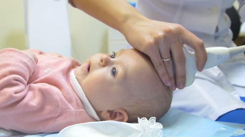
How is an ultrasound performed on infants?
An ultrasound examination is carried out through the child’s non-overgrown fontanel. Therefore, after a year, when the bones of the skull grow together, a computed tomography or MRI is prescribed.
The procedure is carried out according to the following algorithm:
- Treatment of the spring with a special gel that promotes the penetration of ultrasonic rays.
- Setting up the device based on the age of the child being examined.
- Brain examination and recording of results.
Based on the presented conclusion, you should not make a diagnosis yourself. After studying the results, examining the child, and recording concomitant signs of brain development disorders, treatment will be prescribed.
Interpretation of ultrasound results
The results are interpreted by the attending physician; sometimes a consultation with a neurosurgeon is required. If the examination reveals that the baby’s ventricles are dilated, but there are no pathological symptoms, it is necessary to undergo the examination again.
In addition to the size and depth of the elements of the liquor apparatus, which were mentioned above, the following indicators are provided: the interhemispheric fissure should be no more than 3 mm;
subarachnoid space about 3mm.
These measurements indicate the condition of the ventricles and the degree of dilatation. If they are greatly enlarged, there are disturbances in the structures of the brain. The lateral ventricles should not exceed 4 mm, otherwise hydrocephalus is diagnosed.
Treatment of the disease
Dilatation therapy includes medication and physical therapy.
To treat dilation of the lateral and azygos ventricles of the brain in newborns, the following are prescribed: diuretics to reduce the production of cerebrospinal fluid; nootropics to improve blood circulation; central nervous system sedatives; gymnastics and massage of the child to improve the child’s condition and relieve muscle tone; vitamin complexes to prevent rickets.
If the enlargement of the ventricles is a consequence of an infectious disease, antibiotics and antiviral drugs are prescribed. In case of violations of the integrity of the skull and brain, surgical treatment is performed.
Consequences and complications
The consequences of an enlarged cerebral ventricle can be different. It all depends on the degree of expansion and localization of the pathology. The main complications that can occur if medical recommendations are not followed:
- loss of vision and hearing;
- impaired coordination, lack of physical and mental activity;
- lagging behind peers;
- paralysis;
- constant growth of the head, deformation of the bones of the skull;
- epileptic seizures and loss of consciousness;
- hallucinations;
- hemorrhagic shock;
- paralysis;
- death.
If an ultrasound reveals a slight enlargement of the ventricles, but the baby is not capricious and develops according to the norm, a repeat examination is scheduled. To avoid the development of possible complications, do not ignore doctor’s orders. Complete all necessary examinations and treat your child.
The ventricles of the brain are considered an anatomically important structure. They are presented in the form of peculiar voids, lined with ependyma and communicating with each other. During development, brain vesicles are formed from the neural tube, which are subsequently transformed into the ventricular system.
Tasks
The main function performed by the ventricles of the brain is the production and circulation of cerebrospinal fluid. It provides protection to the main parts of the nervous system from a variety of mechanical damage, maintaining a normal level of cerebrospinal fluid takes part in the delivery of nutrients to neurons from the circulating blood.
Structure
All ventricles of the brain have special choroid plexuses. They produce liquor. The ventricles of the brain are connected to each other by the subarachnoid space. Thanks to this, the movement of cerebrospinal fluid occurs. First, from the lateral ones it penetrates into the 3rd ventricle of the brain, and then into the fourth. At the final stage of circulation, the outflow of cerebrospinal fluid into the venous sinuses occurs through granulations in the arachnoid membrane. All parts of the ventricular system communicate with each other using channels and openings.

Kinds
The lateral sections of the system are located in the cerebral hemispheres. Each lateral ventricle of the brain communicates with the cavity of the third through a special foramen of Monroe. The third section is located in the center. Its walls form the hypothalamus and thalamus. The third and fourth ventricles are connected to each other through a long canal. It is called Sylvian Passage. Through it, cerebrospinal fluid circulates between the spinal cord and brain.
Lateral divisions
Conventionally, they are called the first and second. Each lateral ventricle of the brain includes three horns and a central section. The latter is located in the parietal lobe. The anterior horn is located in the frontal, the lower - in the temporal, and the posterior - in the occipital zone. In their perimeter there is a choroid plexus, which is distributed quite unevenly. So, for example, it is absent in the posterior and anterior horns. The choroid plexus begins directly in the central zone, gradually descending into the lower horn. It is in this area that the size of the plexus reaches its maximum value. For this reason, this area is called a tangle. Asymmetry of the lateral ventricles of the brain is caused by a disturbance in the stroma of the tangles. This area is also often subject to degenerative changes. This kind of pathology is quite easily detected on ordinary radiographs and carries a special diagnostic value.

Third cavity of the system
This ventricle is located in the diencephalon. It connects the lateral sections with the fourth. As in other ventricles, the third contains choroid plexuses. They are distributed along its roof. The ventricle is filled with cerebrospinal fluid. In this department, the hypothalamic groove is of particular importance. Anatomically, it is the border between the visual thalamus and the subtubercular region. The third and fourth ventricles of the brain are connected by the aqueduct of Sylvius. This element is considered one of the important components of the midbrain.
Fourth cavity
This section is located between the pons, cerebellum and medulla oblongata. The shape of the cavity is similar to a pyramid. The floor of the ventricle is called the rhomboid fossa. This is due to the fact that anatomically it is a depression that looks like a diamond. It is lined with gray matter with a large number of tubercles and depressions. The roof of the cavity is formed by the lower and upper brain sails. It seems to hang over the hole. The choroid plexus is relatively autonomous. It includes two lateral and medial sections. The choroid plexus attaches to the lower lateral surfaces of the cavity, extending to its lateral inversions. Through the medial foramen of Magendie and the symmetrical lateral foramina of Luschka, the ventricular system communicates with the subarachnoid and subarachnoid spaces.

Changes in structure
The expansion of the ventricles of the brain negatively affects the activity of the nervous system. Their condition can be assessed using diagnostic methods. For example, a computed tomography scan reveals whether the ventricles of the brain are enlarged or not. MRI is also used for diagnostic purposes. Asymmetry of the lateral ventricles of the brain or other disorders can be caused by various reasons. Among the most popular provoking factors, experts call increased formation of cerebrospinal fluid. This phenomenon accompanies inflammation in the choroid plexus or papilloma. Asymmetry of the ventricles of the brain or changes in the size of the cavities may be a consequence of impaired outflow of cerebrospinal fluid. This happens when the holes of Luschka and Magendie become impassable due to the appearance of inflammation in the membranes - meningitis. The cause of obstruction may also be metabolic reactions due to venous thrombosis or subarachnoid hemorrhage. Often, asymmetry of the ventricles of the brain is detected in the presence of space-occupying neoplasms in the cranial cavity. This could be an abscess, hematoma, cyst or tumor.

General mechanism for the development of disturbances in the activity of cavities
At the first stage, there is difficulty in the outflow of cerebral fluid into the subarachnoid space from the ventricles. This provokes expansion of the cavities. At the same time, compression of the surrounding tissue occurs. Due to the primary blockage of fluid outflow, a number of complications arise. One of the main ones is the occurrence of hydrocephalus. Patients complain of sudden headaches, nausea, and in some cases vomiting. Disorders of autonomic functions are also detected. These symptoms are caused by an acute increase in pressure inside the ventricles, which is characteristic of some pathologies of the liquor-conducting system.

Cerebral fluid
The spinal cord, like the brain, is suspended inside the bone elements. Both are washed with liquor from all sides. Cerebrospinal fluid is produced in the choroid plexuses of all ventricles. The circulation of cerebrospinal fluid is carried out due to the connections between the cavities in the subarachnoid space. In children, it also passes through the central spinal canal (in adults, it becomes overgrown in some areas).


 Brief description of the characters Heart of a Dog")


